

What is microsurgical varicocelectomy?
Varicocele surgeries performed under microscopes are called microscopic varicocelectomy.
Today, varicocele surgeries performed using the microsurgical method are accepted as the gold standard method for treating varicocele. The surgery is carried out through a two-centimeter incision in the inguinal region. All enlarged veins of the testicle are ligated under the microscope. The advantage of using a microscope is that it allows seeing all veins and protecting the arteries thanks to the increased visibility. With the microsurgical varicocelectomy operation performed with experienced hands, the possibility of varicocele recurrence is reduced, and the risks associated with the operation are minimized.
What are the symptoms of varicocele?
Varicocele generally does not show any symptoms. But, it may sometimes cause pain in the testicle.
The intensity of the varicocele pain increases with physical exercises and standing for long times. It tends to increase later in the day and decreases when you lie down and rest. Varicocele progresses and becomes evident in the scrotum over time.
What causes varicocele?
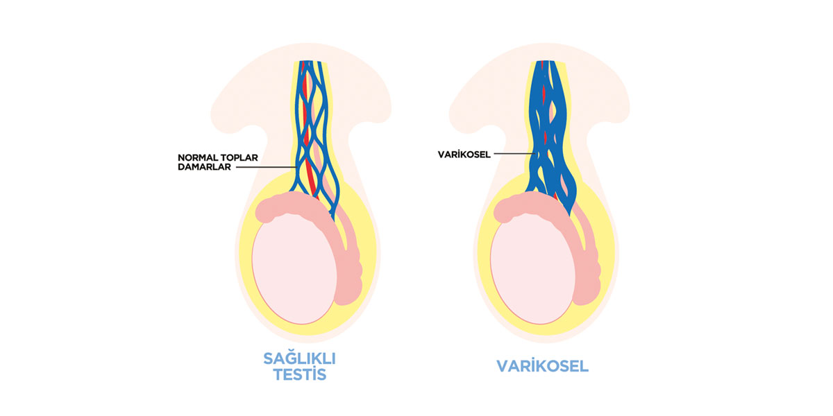
Although there is no clearly defined reason for varicocele, it is thought to be caused by the malfunction of the valves in the veins of the testicle. As a result of the malfunction of these valves, the veins enlarge, and the venous blood accumulates in and around the testicle, which may lead to poor sperm quality or count.
Due to the position of the left testicle vein, varicocele is generally seen on the left side. However, it is known that unilateral varicocele may disrupt sperm production in both testicles.